First! Lets looks at these pearly whites! This is an x-ray of normal teeth!
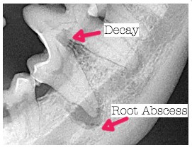
This xray shows the large tooth on the bottom. An abcess has formed at the bottom of the left root and an abcess is forming on the right root. This tooth required extraction!
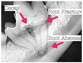
This xray shows the large tooth on the bottom jaw. The roots are slanted with an abscess forming at the bottom. In addition, the root on the right is fractured! There is also some decay in between the roots. This tooth required extraction!
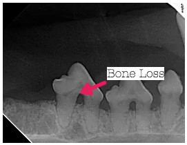
This xray shows some bone loss. Normally the bone will hug right up into the bifurcation (see the normal xray). This is a result of severe dental disease and tartar. These teeth will need extraction.
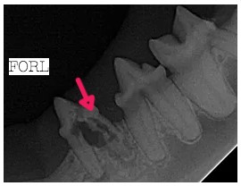
This xray shows a Feline Oral Resporptive Lesion (FORL). This is a result of the body attacking the tooth and it is not completely known why this occurs. They appear as red/pink areas on the tooth itself and can be VERY painful.
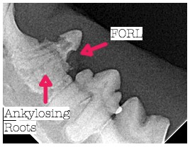
This x-ray shows another FORL. Also the roots are starting to turn to bone (Ankylosing). Notice that the roots are not well defined like in the normal xray.